Awards
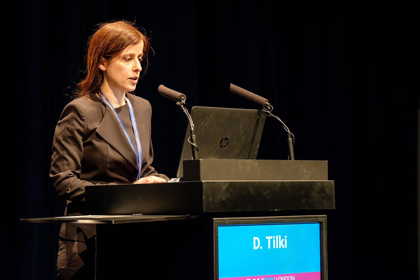
Our visiting faculty member Prof. Derya Tilki received EAU 2020 Crystal Matula Award.

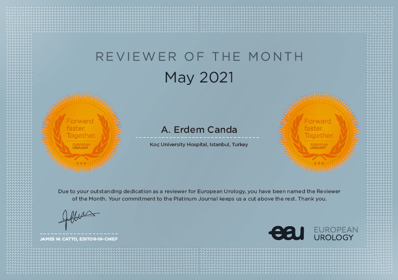
Prof. A. Erdem Canda from our department was nominated as '' Reviewer of the month of European Urology May 2021 issue'' and his name together with Koç University Hospital appears at the main website of this journal that is the urology journal with the highest impact factor.
PDF Download
PDF Dosyası

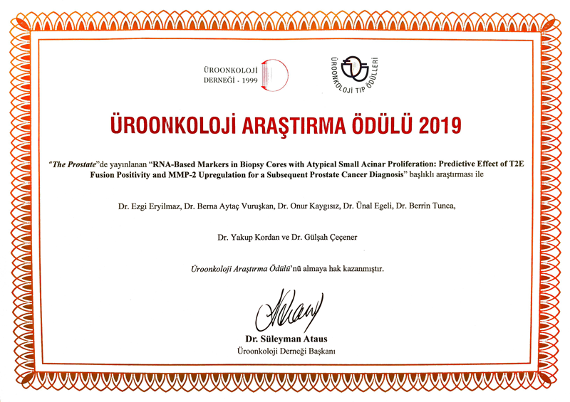
Urooncology 2019 Scientific Study Award
Prof. Yakup Kordan who took part and co-authored in the study entitled ”RNA based markers in biopsy cores with atypical small acinar proliferation: Predictive effect of T2E fusion positivity and MMP-2 upregulation for a subsequent prostate cancer diagnosis” has received Urooncology 2019 Scientific Study Award that was also published in The Prostate journal.
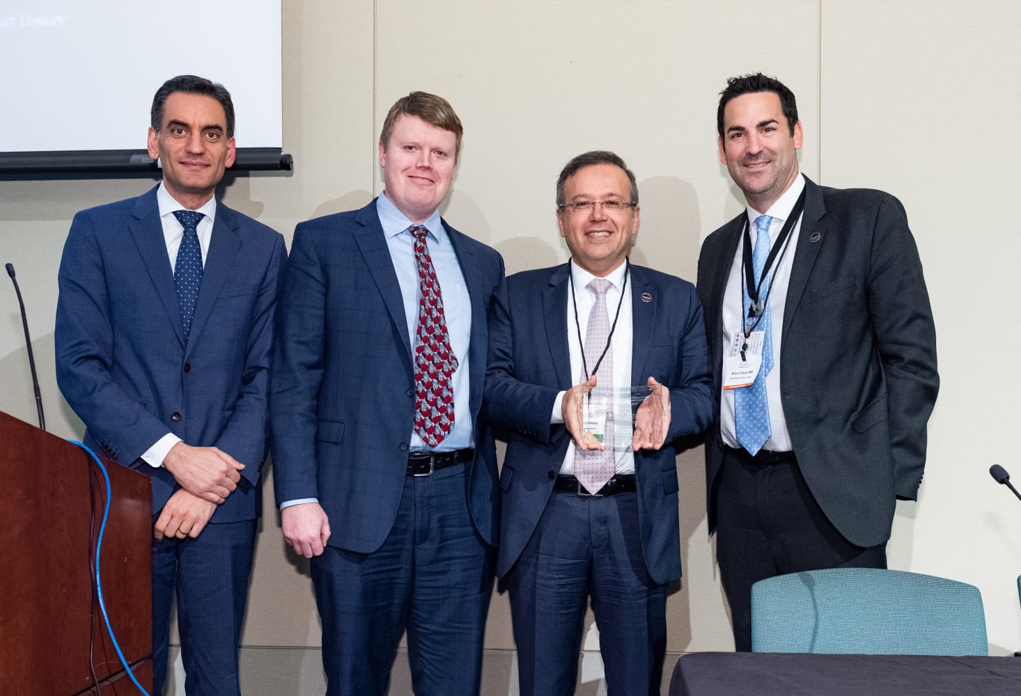
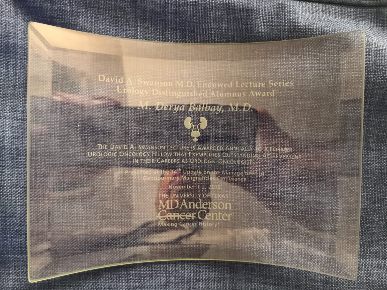
1-2 November 2019 Update on the management of Genitourinary Malignancies, Houston, Texas, USA
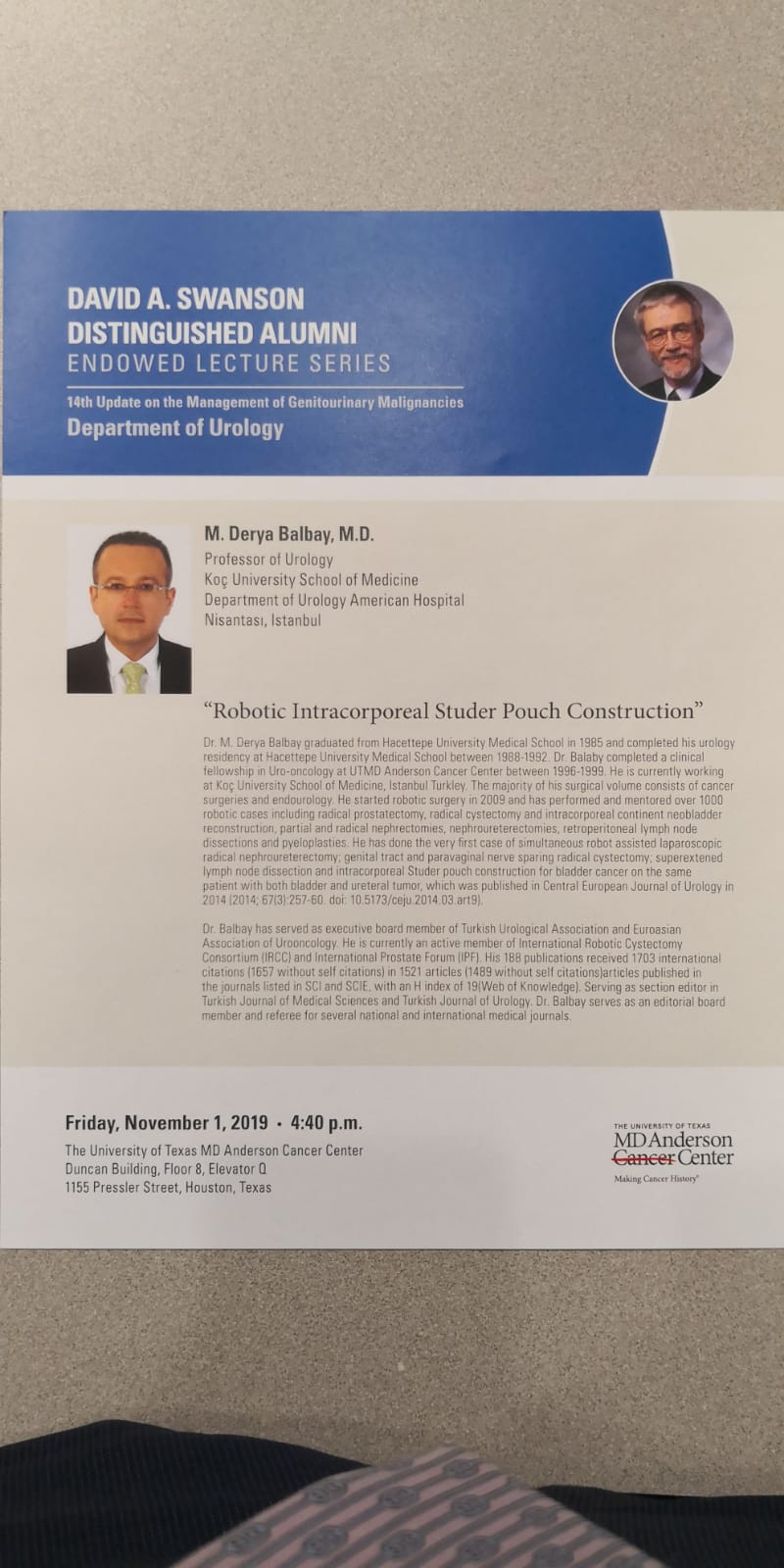
Prof. M. Derya Balbay received David A. Swanson Distinguished Alumnus Award who was an invited speaker and gave a talk on his own surgical technique ”Robotic intracorporeal Studer pouch formation by using Balbay’s technique following robotic radical cystectomy for bladder cancer” during the David A. Swanson Distinguished Alumni Lectures session.
Balbay MD, Canda AE, Kiremit MC, Koseoglu E. Intracorporeal Studer pouch formation with Balbay’s technique following robotic radical cystectomy for bladder cancer: experience with 22 cases with oncologic and functional outcomes. 2019; November, J Endourol, accepted for publication.

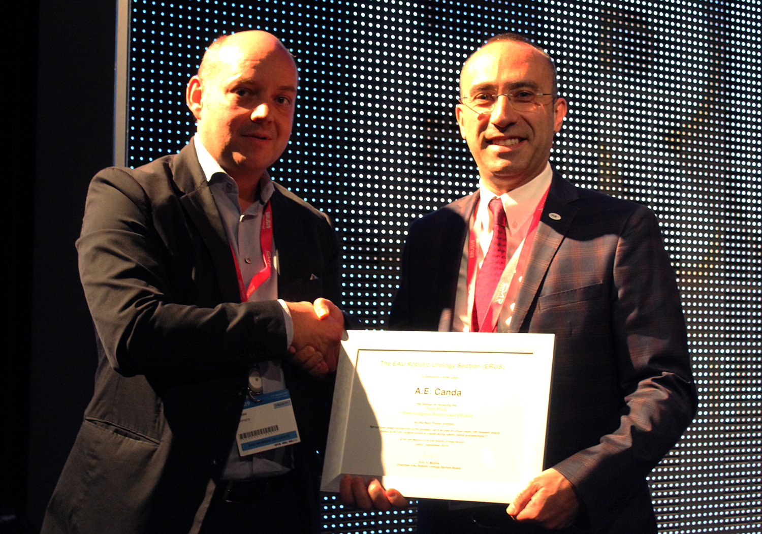
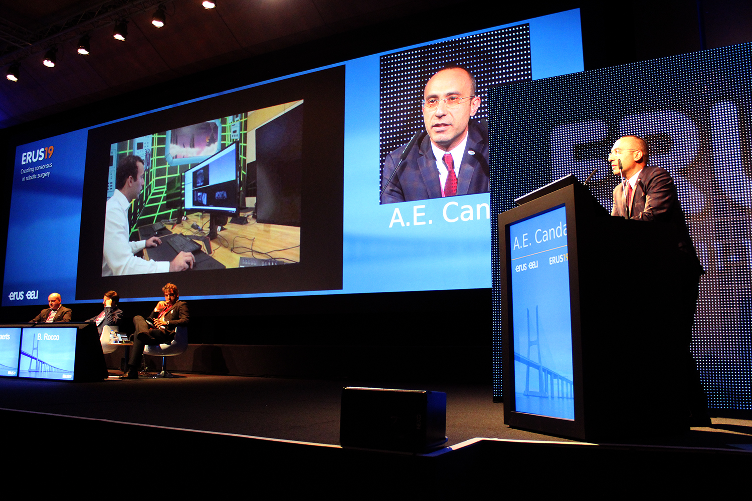
16th Meeting of the EAU Robotic Urology Section (ERUS19), 11-13.September.2019, Lisbon, Portugal
Our study received 3rd prize poster award during the 16th Meeting of the EAU Robotic Urology Section (ERUS19).
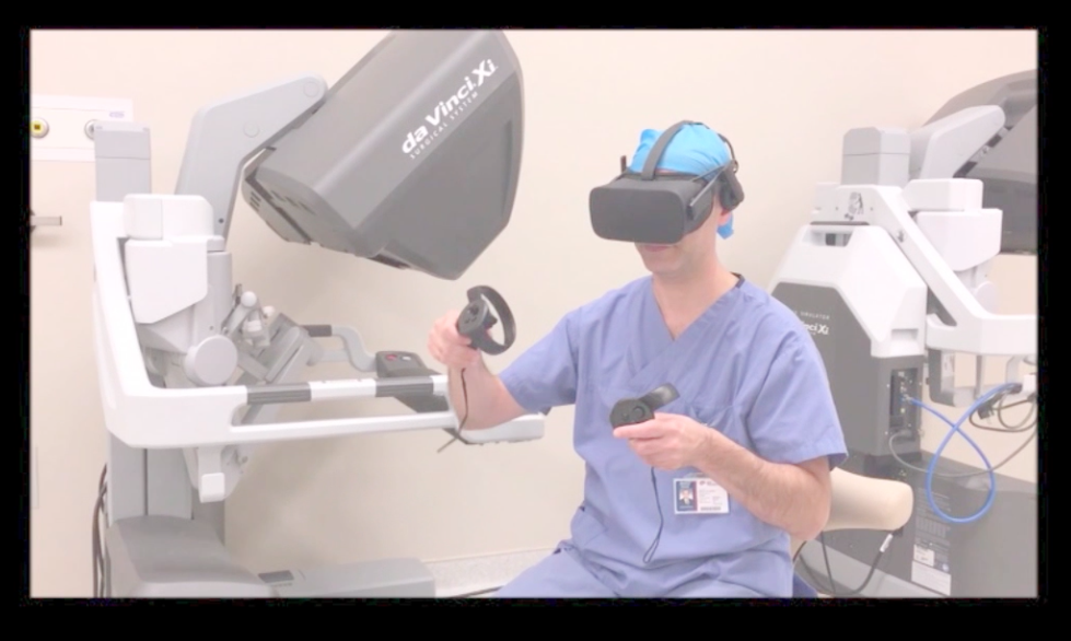
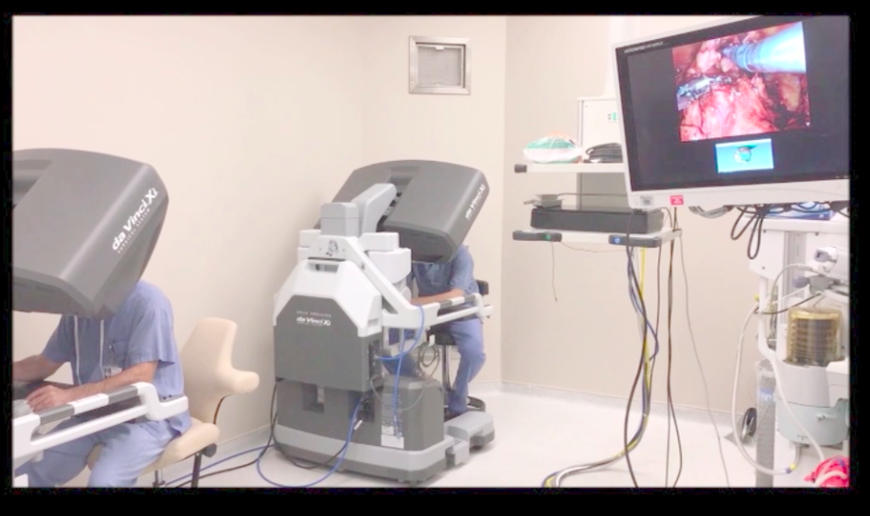
3D real time image reproduction of the prostate: Can it be used on virtual reality (VR) headsets and/or tilepro of Da Vinci surgical system as a guide during robotic radical prostatectomy?
Sertac Fatih Aksoy1, Emre Altinmakas2, Abdullah Erdem Canda3, Ersin Koseoglu3, Tarik Esen3,4
1Sabancı University, Collaboration Space, Istanbul, Türkiye
2Koç University, School of Medicine, Department of Radiology, Istanbul, Türkiye
3Koç University, School of Medicine, Department of Urology, Istanbul, Türkiye
4VKF American Hospital, Department of Urology, Istanbul, Türkiye
Objective: To create 3D prostate images that can be transferred to virtual reality (VR) headsets and tilepro of Da Vinci surgical system.
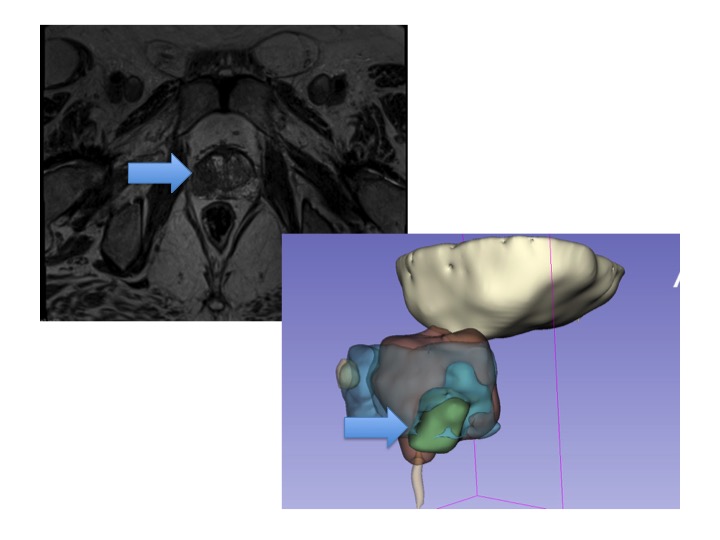
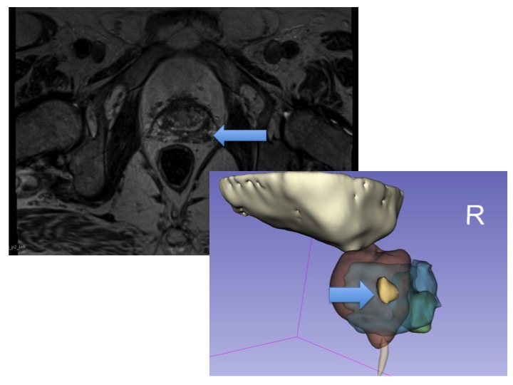
Methods: Axial T2W 3D-TSE sequence was used in multiparametric prostate magnetic resonance imaging (MRI). Borders of the tumor(s) and anatomical structures were marked by the uro-radiologist. In order to obtain the accurate segmentation of the tumor(s) and surrounding anatomical structures, interactive medical image segmentation methods were used such as, fast growcut algorithm and morphological contour interpolation. Simple region growing techniques were used after primary applications for improving the results of the initial methods. Urinary bladder, prostate (peripheral zone was separated) and urethra were segmented to give a better understanding.
Results: Tumor 1 (Figure 1). A 2 cm markedly hypointense mass arising from the right peripheral zone of the midgland is seen on axial T2W images. It shows restricted diffusion on DWI and ADC map. Mean ADC value is 570×10-6mm2/s, PIRADS5. Tumor 2 (Figure 2). A 0.8 cm lesion is noted at the left base of prostate gland. It shows markedly hypointense signal on T2W images and restricted diffusion on DWI and ADC map. Mean ADC value is 760×10-6 mm2/s, PIRADS4. 3D images were constructed and were transferred to VR headsets. Tumor 1 had 851.781 mm2 surface area, 1762.45 mm3 and 1.76 cm3 volumes. Tumor 2 had 129.258 mm2 surface area, 123.931 mm3 and 0.12 cm3 volumes.
Conclusion: 3D image representation of the prostate and transfer of the images on VR headsets and/or tilepro of Da Vinci surgical system might be used as a promising tool to help the operating console surgeon to better understand the size, location and characteristics of the tumor(s) and the prostate. It might be used as a guide during robotic radical prostatectomy and might have an impact on the learning curve and oncologic outcomes.