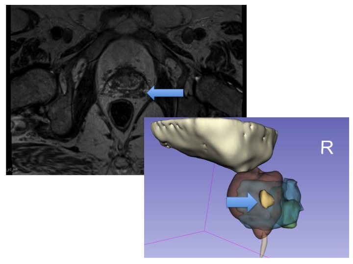
Methods: Axial T2W 3D-TSE sequence was used in multiparametric prostate magnetic resonance imaging (MRI). Borders of the tumor(s) and anatomical structures were marked by the uro-radiologist. In order to obtain the accurate segmentation of the tumor(s) and surrounding anatomical structures, interactive medical image segmentation methods were used such as, fast growcut algorithm and morphological contour interpolation. Simple region growing techniques were used after primary applications for improving the results of the initial methods. Urinary bladder, prostate (peripheral zone was separated) and urethra were segmented to give a better understanding.
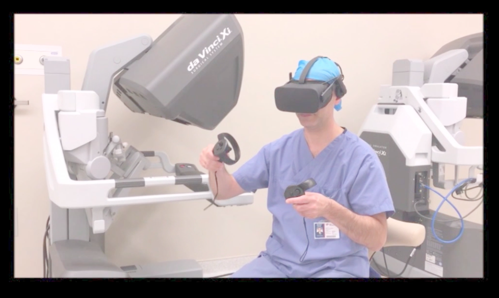
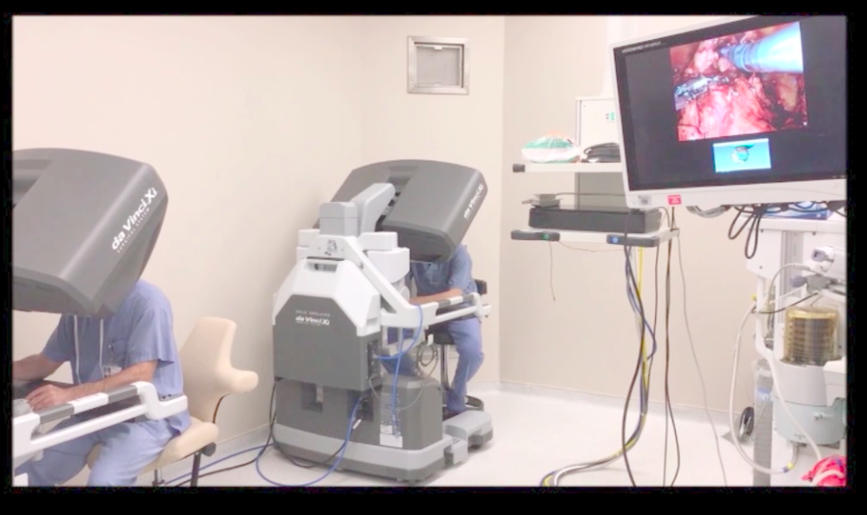
Results: Tumor 1 (Figure 1). A 2 cm markedly hypointense mass arising from the right peripheral zone of the midgland is seen on axial T2W images. It shows restricted diffusion on DWI and ADC map. Mean ADC value is 570×10-6mm2/s, PIRADS5. Tumor 2 (Figure 2). A 0.8 cm lesion is noted at the left base of prostate gland. It shows markedly hypointense signal on T2W images and restricted diffusion on DWI and ADC map. Mean ADC value is 760×10-6 mm2/s, PIRADS4. 3D images were constructed and were transferred to VR headsets. Tumor 1 had 851.781 mm2 surface area, 1762.45 mm3 and 1.76 cm3 volumes. Tumor 2 had 129.258 mm2 surface area, 123.931 mm3 and 0.12 cm3 volumes.
Conclusion: 3D image representation of the prostate and transfer of the images on VR headsets and/or tilepro of Da Vinci surgical system might be used as a promising tool to help the operating console surgeon to better understand the size, location and characteristics of the tumor(s) and the prostate. It might be used as a guide during robotic radical prostatectomy and might have an impact on the learning curve and oncologic outcomes.
